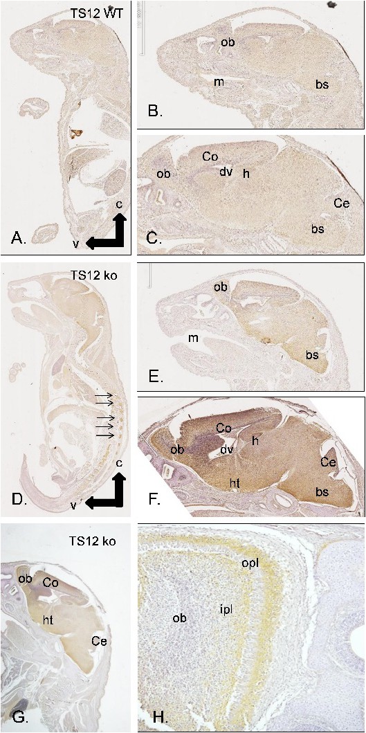
Fig. 5. Immunohistochemical analysis of neurocan expression in neonatal mouse brain. Complete parasagittal sections of a wild-type mouse (wt; A) and an Adamts12-deficient mouse (TS12 ko; D). An immunoreaction was only detected in Adamts12-deficient mice (D-H). m: mouth; ob: olfactory bulb; Ce: cerebellum; Co: cortex; h: hippocampus; ht: hypothalamus; bs: brain stem; dv: diencephalic vector; c: cephalic; v: ventral; opl: outer plexiform layer; ipl: internal plexiform layer. The arrows indicate an immunoreaction for neurocan in the rachidial ganglia of the spinal nerves.